
At her Vancouver office, professor Claudia Krebs looks at illustrations in a 1967 edition of an anatomy atlas originally edited by Werner Spalteholz and taken over by Rudolf Spanner. These are reproductions that appeared in the original Spalteholz atlas, and are not problematic. On her computer are examples of how anatomical images (in this case, images which do not have a Nazi history) are being reused.Photography by Tijana Martin/ The Globe and Mail
Claudia Krebs filled out the online form and arrived at the University of British Columbia library, masked, to pick up her order. She hauled the old anatomy atlases home in plastic bags. It was 2021, the university was still in COVID lockdown, and she was preparing to teach a class on ethics in biomedical visualization. Pernkopf was the natural place to start.
Pernkopf’s Atlas of Topographic and Applied Human Anatomy was once the gold standard in illustrated anatomy texts. Originally published in 1937, it was the first book of its kind to use full-colour offset printing. The quality of the paintings was without equal, and the cross-sectioned depictions of the body were displayed in helpfully layered dissection steps.
Translated from German into English and other languages, it was found in medical schools internationally. For decades, it was an essential resource for everyone from med students learning their way around the human body to veteran surgeons. “There was no other atlas like it in the world,” says William Seidelman, emeritus professor in the department of family and community medicine at the University of Toronto.

Eduard Pernkopf's atlas has been out of print for decades, but as Dr. Krebs demonstrated, its illustrations have had a long afterlife.
Now, it’s a pariah.
Dr. Seidelman, 83, is one of the key figures responsible for investigating and exposing how the bodies in the Pernkopf atlas were procured: they were victims of the Nazis. The issue came to a head in the 1990s and the book went out of print – although ethical questions around Pernkopf still swirl.
But then, in another stack of old anatomy atlases Dr. Krebs subsequently checked out, she made a shocking discovery. The Pernkopf isn’t the only anatomy book with this nefarious history. She found another one at the UBC library, thick with images of Nazi victims.
There was something else, too, about this other atlas. It included details Pernkopf’s did not: biographical information, however scant, about the drawn victims, clues which would set Dr. Krebs onto the investigation of a lifetime as she attempted to find out their identities. “It’s somewhat shocking that after 80 years, it took me just going to the library for this to come to light,” says Dr. Krebs, Professor of Teaching in the Department of Cellular and Physiological Sciences at UBC’s Faculty of Medicine.
In her investigation, she also discovered copious reproductions of Pernkopf illustrations in other resources. They have been redrawn, copied, traced, flipped – and still populate important medical journals and texts. “We’re all using these images in our lectures that are traced or inspired by this Pernkopf atlas, that are not as original as we thought they were,” explains Dr. Krebs, a German professor who lives in Vancouver.
Even if the use is often unwitting, this new information raises old ethical questions that emerged with the Pernkopf discovery decades ago. These images have benefited countless medical professionals and their patients. But should they still be used?
Dr. Krebs presented her work publicly for the first time at an anatomy conference late last month in Toronto, to stunned silence in the room. Afterward, she shared what she believes could be the potential impact. “I am not exaggerating when I say that this symposium may have changed the way we deal with anatomical illustrations permanently.”

At a lecture hall draped with swastikas and a portrait of Hitler, Eduard Pernkopf addresses the University of Vienna faculty in 1938, when he became the new dean of medicine. Austria had just been annexed by Nazi Germany, whose policies of racism and eugenics would permeate Austrian academia.Austrian National Library
Eduard Pernkopf, born in 1888, was an Austrian physician and anatomist – and a devoted Nazi. He was installed as dean of the medical school at the University of Vienna in 1938, after the Anschluss.
There’s an infamous photo of Pernkopf’s first address to his faculty. He is standing at the lectern in his Nazi uniform, surrounded by medical school colleagues giving him the Hitler salute. In that address, he told the physicians they had a responsibility in “eliminating the unfit and defective.”
Between 1938 and 1945, some 1,377 bodies made their way from sites of execution to Pernkopf’s lab. They were, for the most part, political prisoners and people the Nazis considered common criminals in their reign of terror, often beheaded by guillotine by the Gestapo. These are the bodies used in his famous atlas. The illustrator’s signatures sometimes incorporated Nazi iconography, including swastikas and SS runes – crudely removed in later English editions.
In addition to educating students about the history of the Pernkopf atlas, Dr. Krebs also wanted to explore its antecedents. Who was inspired by whom? What inspired Pernkopf himself? With this in mind, she was interested in an atlas originally edited by German anatomist Werner Spalteholz, published in three volumes at the turn of the last century – long before the Nazis were around to taint the process.
In their discussions about the issue, Dr. Sabine Hildebrandt, an anatomy educator at Harvard Medical School who studies the history and ethics of anatomy, mentioned that there were postwar editions of the Spalteholz atlas that included some Pernkopf drawings: a 15th edition published in 1954, and a 16th in 1961.
After Spalteholz’s death in 1940, these new versions were edited by another German physician, Rudolf Spanner, who was the director of the Institute of Anatomy in Danzig – the German name for Gdansk, Poland – from 1939 to 1945.
One of the credits in a 16th-edition Spalteholz atlas that mentions Danzig, now Gdansk, Poland.
Dr. Krebs was able to find a copy of the 16th edition at the UBC library, published in English in 1967. And she did indeed encounter some Pernkopf drawings.
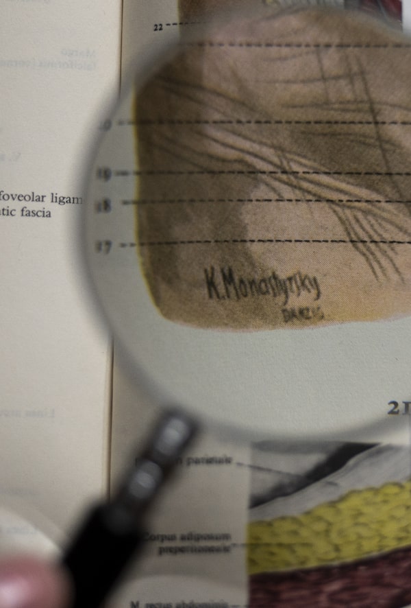
But Dr. Krebs was so familiar with the previous edition of the atlas that she was able to spot something else: new illustrations, not from the Pernkopf atlas, and not in the earlier Spalteholz book either. The artist signatures, sometimes embedded into organs and tissue and visible only through a magnifying glass, indicated where and when they were drawn. Many had been made in “Danzig.” Also included, she realized with creeping shock, were the years they were drawn: 1942 through 1945.
“All of a sudden, I could place the creation of these images in Nazi-occupied Poland,” Dr. Krebs recalls. “My heart dropped.”
Further, dozens of the illustrations had typed captions listing some details about who was depicted – identifications such as 33-year-old man, 37-year-old female, “fresh 18-year-old subject.”
“It was horrible,” she says, noting that the first signature from wartime she spotted was on a drawing of a 12-year-old boy. “It was just like being punched in the gut.”
The 12-year-old boy is drawn in such a way that the entire core of his body is visible, looking in from the front of his abdominal cavity – so his kidneys and blood vessels can be seen. In another drawing, the same boy’s thorax and abdomen are shown together. Another illustration depicts an emaciated 18-year-old boy who is circumcised – so likely Jewish.

The two postwar editions of the Spalteholz atlas were edited by Rudolf Spanner, a Nazi. Some of his contributions were shocking, as Dr. Krebs discovered.
Dr. Krebs then began her deep dive into where these bodies came from – and who ran the lab where they had ended up.
Rudolf Spanner was already a notorious doctor figure from the Nazi era – but not because of anatomical illustrations. Born in 1895 in Koblenz, Germany, Spanner was a Nazi party member tasked with establishing and expanding the anatomy department at the Danzig Anatomical Institute during the Second World War. In 1939, he was nominated for the Nobel Prize in Physiology or Medicine.
He is well known by Second World War historians for allegations that he’d used his lab to make soap from human bodies.
In Gdansk, a controversial plaque memorializes this. In four languages, it explains: “In this building, during World War II the Nazis used the bodies of victims of genocide … as material to produce soap. People brought this fate upon people.”
The allegations against Spanner were never proven and likely did not happen on any sort of industrial scale, according to academic and scientific investigations, including the comprehensive book Soap from Human Fat: The Case of Professor Spanner by Monika Tomkiewicz and Piotr Semkow.
Spanner was indicted in Nuremberg for the soap allegations, but never tried. He later received what’s known as class five denazification status – fully exonerated of any Nazi deeds. He continued to practice medicine after the war and was appointed director of the anatomy department at the University of Cologne in 1949.
The University of Cologne is where Dr. Krebs, who was born in the city, attended medical school. When she began this investigation, and first came across Spanner’s photograph, she recognized it: at university, she had often passed by the same photograph of Spanner, the Nobel Prize nominee, on the wall.
Two editions of Spanner’s Spalteholz atlas were published – the second in 1961, a year after Spanner died. The Spalteholz-Spanner Atlas of Human Anatomy was widely used until about 1970. If anybody noticed the alarming dates and locations noted in the signatures, or put two-and-two together to figure out where these bodies came from, they didn’t say anything.
As Dr. Krebs learned from Soap from Human Fat, the bodies arrived at Spanner’s lab from Gestapo execution sites in Danzig, Posen and Konigsberg; the Danzig prison; the Konradstein Psychiatric Institution; and the Stutthoff concentration camp.
While documents naming the Nazi victims taken to Pernkopf’s lab were destroyed toward the end of the war, some lists did survive of those brought to Spanner’s Danzig lab.
Dr. Krebs pulled out all the illustrations that had biographical data and checked them against those in the original version of the Spalteholz atlas. Anything that wasn’t in the earlier version was subject to investigation. Even the most minimal biographical data was a clue.
“And so we went through all of these and tried to find: who are these people?”

A Polish defendant, shackled in the background, is tried at a Nazi special court in Bydgoszcz (Bromberg, in German). Poland's occupiers used such courts, called Sondergerichte, to summarily execute prisoners for nearly any reason. Some of their bodies would end up in Spanner’s lab.Alamy
Last fall, three fourth-year UBC medical students signed on to help Dr. Krebs investigate as part of their course work. For six weeks, they met daily at UBC’s Life Sciences Centre in a room set aside for their work. For hours each day they sat at their laptops, surrounded by the Spanner atlas and other textbooks.
They cross-referenced the identifying demographics in the atlas with other sources, including a treasure trove Dr. Krebs found at the back of Soap from Human Fat: lists of names of victims whose bodies were brought to the Danzig Anatomical Institute from Gestapo execution sites and the psychiatric hospital.
The students also consulted international archival databases available online, including those from Germany’s Arolsen Archives, Yad Vashem’s database of Shoah Victims’ Names in Israel, and the Institute of National Remembrance in Poland.
“There were moments when you just feel overwhelmed,” said student Katerina Schwab.
The team created a database with all of the names, birth and death dates, how they were executed, where they were imprisoned – any information they could get their hands on so it could be cross-referenced.
Although the biographical descriptions on the drawings were minimal, connections were made.
In one case, a caption indicated the illustration depicted a body belonging to a 33-year-old male. Cross-referencing the lists of victims sent to the institute from the Gdansk prison, the group found two males of that age. One was killed in 1942, the other in 1944. The illustration was signed 1943. They had their man – probably.
“You kind of sit there, you look at their name,” said student Ivica Bratanovic about another illustration. “Wow, we figured out who this person was, who was so unknown. But that hand – some of them were drawings of dissections of hand – has been nameless and seen by thousands of med students. And now there’s a name to that.”
Mr. Bratanovic, who will officially be Dr. Bratanovic next month, is going into radiology, an anatomy-heavy specialty. “Every time I use those textbooks, paying tribute is really important,” he says.

Dr. Krebs was shocked to find new Nazi-era anatomical illustrations – and descriptions of some of the subjects.
With all of this new information, Dr. Krebs had to tell the world. She has given many talks over the years – but revealing her new discoveries in public for the first time, at last month’s American Association for Anatomy conference in Toronto, was the hardest of her career.
She began by showing how illustrations in publications still in print originated from Pernkopf, demonstrating how they have been copied or otherwise manipulated and are still very much in use.
Many of the standing-room-only audience of scientists, anatomists, medical illustrators and students gathered for the talk were shocked, she says. “They were like, Oh my God, I’ve used that.”
Then she revealed her Spanner discovery.
Dr. Krebs would first display a slide with the name of the person she and her students identified, as well as records they found in the archive. She recited information about the person out loud: birth and death dates, profession, what they were arrested for – mostly, they were Polish resistance fighters. Often, if the person was married, she was able to access the name of their wife.
She read the information printed on the slides: “Stanislaw Jozef Lewsinki,” she said, adding his dates of birth and execution, and his age, 47. He was a police officer, “married to Zorja.”
Then, in silence, she would show the next image: the page from the atlas, the likely illustration of that person.
“I think it really brought the humanity of the people,” Dr. Krebs said later. “Because when you have the name, and you have a little bit of their story, as sparse as it is, and then you see their hand dissected, it really makes you question: where do your images come from? Who consented or didn’t consent? What is the cost, the human cost of the knowledge that we have?”

Dr. Krebs demonstrates three pictures of the anterior thigh from a different 19th-century atlas. She believes other illustrations – ones that used victims of Nazis – should not be used. ‘We can draw new pictures. We don’t have to trace those.’
There’s a well-known story in anatomy ethics circles about the Pernkopf atlas, which exemplifies the ethical dilemma created by the availability of these images.
A patient, experiencing excruciating pain in her knee, begged her doctor to amputate, if she couldn’t fix it. The surgeon – a renowned expert – felt she needed to consult the Pernkopf atlas, but didn’t know if she should.
She contacted Joseph Polak, a physician, professor, Chief Justice of the Rabbinical Court of Massachusetts – and child survivor of the Holocaust. He contributed – along with Dr. Hildebrandt, Dr. Seidelman, and others – to the 2017 Vienna Protocol, which addresses what should be done to remains discovered from the Holocaust era.
“She said, ‘The only way I can help this woman, this patient, is by opening the Pernkopf,’” Rabbi Polak recounted during a recent online event hosted by the Medicine, Holocaust and Genocide Studies Program at Cedars-Sinai in Los Angeles.
“What I told her was that if this was going to save an amputation and let another human being live without pain, then just go and get the Pernkopf and look at it,” said Rabbi Polak, who lived in Montreal after the war. “But before you actually do that, gather the patient and her family and tell her about the atlas and tell her where this atlas came from. And who’s in it.”
Although the Pernkopf Atlas has been out of print since the 1990s, some translations were available well into the 2000s, free digital copies of the atlas are available online and used physical copies sell for hundreds of dollars. And the images can be found in other textbooks, including Gray’s Anatomy for Students and the Sobotta Atlas of Anatomy.
“I have probably browsed them and used them without even knowing,” says Mr. Bratanovic.
The 2023 edition of the Sobotta atlas has a new foreword acknowledging the use of the Pernkopf images, co-written by Dr. Hildebrandt and the anatomy editors.
“This decision to reproduce an illustration from the Pernkopf Atlas can only be justified on the basis of a conscious examination of the egregious ethical transgressions of anatomy during National Socialism (Nazi Germany), in memory of the victims of the Nazi regime whose bodies are depicted here,” the new foreword states, in line with Rabbi Polak’s wording from the Vienna Protocol. They are being used to save future patients, it states, in memory of the victims.
Dr. Krebs is pleased that this acknowledgment exists, but she disagrees with the decision to include these pictures. They are part of the history of medical illustration and that’s where they should stay, she believes.
“We can draw new pictures,” she says. “We don’t have to trace those.”

After her research into anatomical atlases, Dr. Krebs disagrees with decisions to keep using images derived from Pernkopf.
Efforts continue to limit the use of the images known to be created from Nazi victims.
The illustration in Gray’s Anatomy was removed. The university in Gdansk has been notified and agrees that the findings need to be shared and publicized.
And when Dr. Krebs returns the Pernkopf atlases to the UBC library, they will go into special collections. “They’ll never leave the library again,” she says, adding that the library has been extremely supportive. Neither will Spanner’s Spalteholz atlas. Historical context will also be provided to borrowers.
Still, these images are everywhere, as she has demonstrated. And now, with artificial intelligence, if computers are being trained to sweep images to create new ones, it seems inevitable that the bodies of Nazi victims will be caught up in this. “The data sources, have they been questioned? Have they been interrogated? Do we know the history of these?” asks Dr. Hildebrandt, author of The Anatomy of Murder: Ethical Transgressions and Anatomical Science during the Third Reich.
Dr. Hildebrandt says the educated need more education in this area: medical illustrators shouldn’t just be able to copy or trace any image without asking where it came from.
Further identification of the victims – perhaps even locating family members – may be possible, however daunting the task. Should families be found, there is the possibility of consulting them on the drawings’ futures.
Of course, identification can never be absolutely certain. And Dr. Hildebrandt, who has reservations about assigning identities of victims to images, thinks that’s an important caveat to keep in mind.
“There is a relatively high likelihood for some of them. I don’t think we can ever go further than that, or whether we should ever go further than that,” she said. “Because we do not want to have the victims remembered by an abstract anatomical painting, even if that should show some of their physique. But I think that’s too dehumanizing … for a person who never agreed to be depicted in this way.”
While Dr. Krebs agrees identification can’t be ironclad, she believes acknowledging what their names likely were is important. “They are part of their personhood, part of their story,” she told the audience in Toronto. But only part of it, she emphasized. “No person can – or should – be reduced to some entries in the administrative ledger of a terror regime.”
After her presentation, attendees lined up to speak to her. An Indigenous woman gave her tobacco, for healing. The little red pouch sits on a shelf in Dr. Krebs’ office, displayed among photos of her children and a slew of teaching awards. It shares space with some handicrafts: a framed uterus made from yarn, and an old-timey needlepoint with a contemporary message about seeking justice: “Nevertheless, she persisted.”
 Marsha Lederman